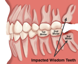
 In most cases, inadequate space in the mouth does not allow the wisdom teeth to erupt properly and become fully functional. When this happens, the tooth can become impacted (stuck) in an undesirable or potentially harmful position. If left untreated, impacted wisdom teeth can contribute to infection, damage to other teeth, and possibly cysts or tumors.
There are several types, or degrees, of impaction based on the actual depth of the teeth within the jaw:
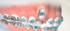 The canine, or eye tooth, normally erupts into the mouth between the ages of 11 and 13. Sometimes one or both canines develop in the wrong position. Often they lie across the roof of the mouth behind the front teeth. If left alone the tooth will not erupt normally and may either damage the roots of the front teeth or push them out of position.
Once the canine is exposed a small bracket is glued to the tooth. Attached to this is a chain which your orthodontist can then use to pull the tooth into the right position. The chain is usually stitched out of the way but it is quite delicate and therefore it is important to be careful when eating for the first few weeks after surgery.
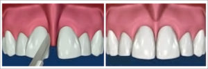
When frenum is attached high or between two upper front teeth, early removal can improve space closure of the front teeth. There is very little post-operative pain and swelling, and the patient can have normal function of the lips and tongue. Many patients hardly notice the effects of the surgery.
The teeth are held firmly in place by strong roots that extend into the jawbone. Molars and premolars tend to have several roots, whereas the front incisors only have a single root. The end or tip of each root is termed the apex. The apex is where the nerves and blood vessels enter the tooth, and aids in the delivery of blood to the crown (the part of the tooth you can see in your mouth).
A root canal treatment refers to the cleaning of the canals and the removal of infected and inflamed tissue within the root. When the inflammation or infection persists after the root canal treatment, an apicoectomy may be required. An apicoectomy is essentially the removal of the apex (or root tip), followed by a filling procedure to seal the root from further infection. When left untreated, infected roots can damage other teeth, spread infection, and cause regression of the jawbone.


A cyst is lined with a kind of tissue called epithelium. This type of tissue normally is found in surface layers, such as the skin and the lining of the mouth. Cysts may form when epithelium cells move into deeper body layers and begin to multiply.
Oral cysts can be found in the jawbone, or in soft tissues such as the salivary glands, skin or inside the mouth.
A cyst is not cancerous but can damage your soft tissue. As the cyst develops, the bone structure around the tooth will feel an extreme amount of pressure. It could eat away from the bone and weaken it.
An oral surgeon usually does this procedure under local anesthesia. If the cyst is infected, you may be given antibiotics.